 Mon Prof Augmenté
The human heart in cutaway: how it works
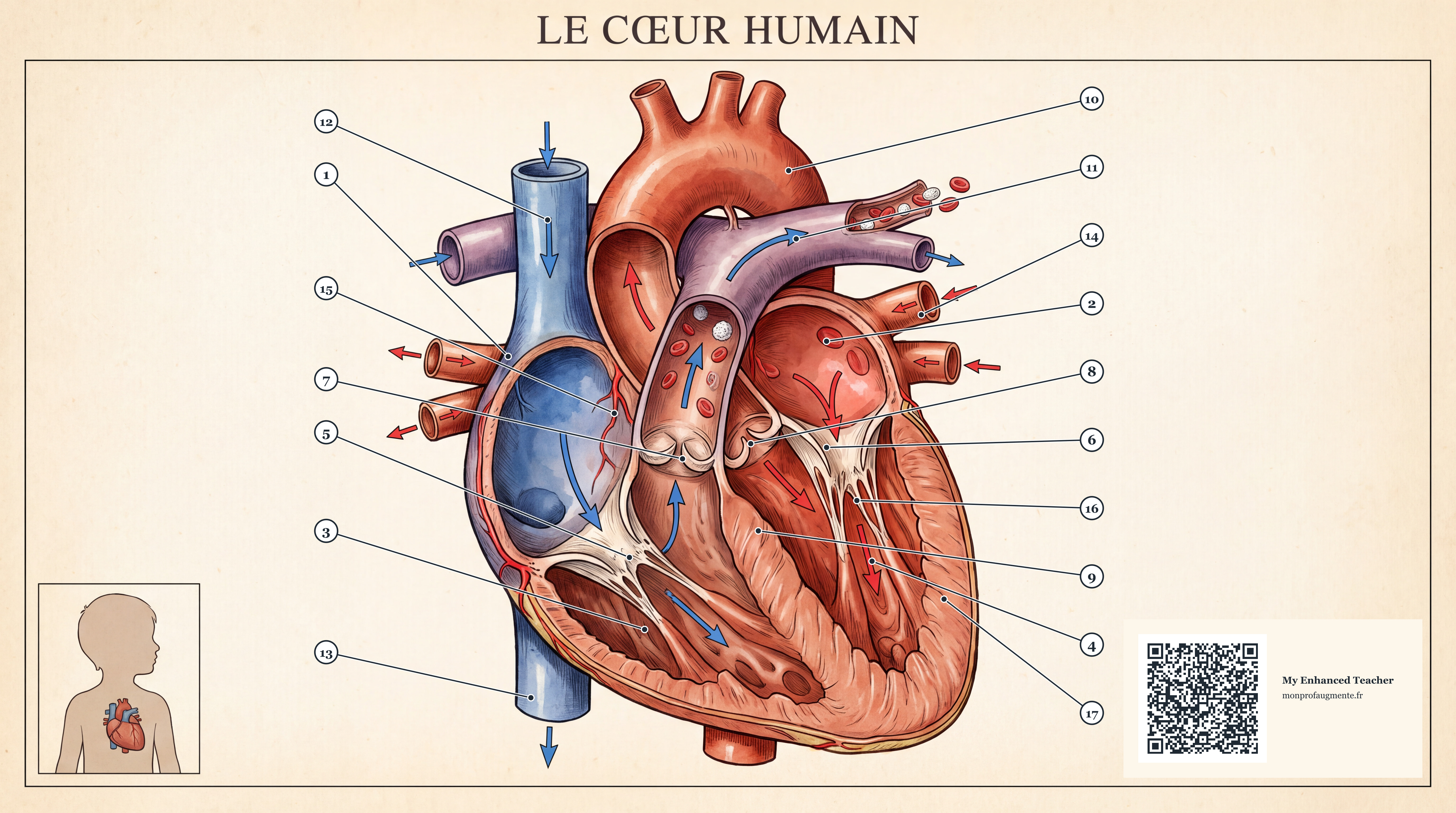
The heart is a muscle about the size of a closed fist that pumps blood around the body without ever stopping. It has four chambers and four valves, and it works as two pumps side by side: the right side sends blood to the lungs, and the left side sends it to the rest of the body. This plate opens the front wall of the heart to show what is inside. Blue is blood low in oxygen, red is blood rich in oxygen. Each number on the picture is explained in the list below.

See it at full size
On this page the picture is scaled to fit your window. The original is 5504×3072 pixels, about 17 megapixels, so there is much more to see than the page shows at once. Three ways to look closer:
- Press “View at full size”. The viewer opens with the image scaled to your window. Choose Actual pixels to show every pixel of the file, one for one, then drag the picture to move around it. Fit returns to the whole plate. Keys: + and − to zoom, 1 for actual pixels, 0 to fit, Esc to close.
- Open the file itself. Open the full-size image in a new tab. Browsers first show it shrunk to the window. Click the image once to switch to 100 % (Chrome, Edge, Firefox and Safari all do this), then scroll or drag to move around.
- On a phone or tablet, open the full-size image the same way and pinch to zoom. The view is sharp because the file really is that large.
Retina and other high-density screens show an image sharper than its pixel count suggests, so “Actual pixels” shrinks the picture on those screens so that one image pixel really is one screen pixel.
Print it
Do not print from the browser window: it prints the page, not the picture at its full size. Use the downloaded file, which contains all 5504×3072 pixels.
- Download the full-resolution JPEG (2.6 MB).
- Open it in Preview (Mac), Photos (Windows) or any image viewer and choose Print. Do not use the browser’s print preview on this page.
- Set the paper to landscape. Choose Fill page or Scale to fit and keep the proportions. The plate is 16:9, so on A-series paper there will be a white band at the top and bottom, and you should not crop it: the QR code is in the corner.
- Choose the highest print quality, and turn off any “draft” or “save ink” mode.
- For a poster, take the same file to a print shop and ask for the largest size you want. Tell them it is 5504×3072 pixels so they know the sharpness they can expect.
| Paper, long side | Sharpness | Good for |
|---|---|---|
| A4 (297 mm wide) | 471 ppi | Sharp on any printer |
| A3 (420 mm wide) | 333 ppi | Best choice: a wall poster you can read up close |
| A2 (594 mm wide) | 235 ppi | Good from arm’s length |
| A1 (841 mm wide) | 166 ppi | Classroom wall, viewed from a distance |
For a sharp print, aim for 150 ppi or more at the distance you will read it from. Print shops usually want 300 ppi for a close-up look; at A3 this plate reaches 333 ppi.
What each number shows
- Right atrium. The right atrium receives blood that is low in oxygen after its trip around the body, through the two large veins called venae cavae. It then passes the blood down into the right ventricle.
- Left atrium. The left atrium receives blood rich in oxygen from the lungs, through the pulmonary veins. It then passes the blood down into the left ventricle.
- Right ventricle. The right ventricle pumps blood to the lungs, where it picks up oxygen. The trip is short, so its wall is thinner than the one on the left.
- Left ventricle. The left ventricle pumps blood rich in oxygen out through the aorta to the whole body. It has to push hard over a long distance, so it has the thickest wall of the four chambers.
- Tricuspid valve. This valve sits between the right atrium and the right ventricle. It has three flaps, called cusps, and lets blood go one way only: forward, never back into the atrium.
- Mitral valve. The mitral valve sits between the left atrium and the left ventricle. It has two cusps and closes when the ventricle squeezes, so blood goes into the aorta and not backwards.
- Pulmonary valve. This valve is at the exit of the right ventricle, at the start of the pulmonary artery. It opens to let blood out toward the lungs and closes to stop it flowing back.
- Aortic valve. The aortic valve is at the exit of the left ventricle, at the start of the aorta. It has three cusps, opens at each beat and then shuts so that blood does not fall back into the heart.
- Septum. The septum is the muscular wall between the left and right sides of the heart. It keeps blood low in oxygen and blood rich in oxygen apart.
- Aorta. The aorta is the largest artery in the body. It leaves the left ventricle, curves over in an arch and sends oxygen-rich blood to the head, the arms and the rest of the body.
- Pulmonary artery. The pulmonary artery leaves the right ventricle and splits in two, one branch for each lung. It is unusual because it carries blood low in oxygen, even though it is an artery.
- Superior vena cava. This large vein brings blood from the head, the neck and the arms back to the right atrium.
- Inferior vena cava. This large vein brings blood from the legs and the belly back up to the right atrium.
- Pulmonary veins. Usually four pulmonary veins carry blood rich in oxygen from the lungs to the left atrium. Their name can be confusing: unlike most veins, they carry oxygen-rich blood.
- Coronary arteries. The heart muscle needs its own supply of blood. The coronary arteries start at the aorta, just above the aortic valve, and run over the surface of the heart to feed the muscle.
- Papillary muscles and chordae. Small muscles called papillary muscles are linked to the valve cusps by thin cords, the chordae tendineae. They hold the cusps in place, like the ropes of a parachute, so that the valves do not flip back.
- Heart muscle (myocardium). The myocardium is the muscle that makes up most of the heart wall. It contracts and relaxes by itself, again and again, to push the blood. It is thickest around the left ventricle.
The human heart at a glance
| Size and weight | About the size of a closed fist; roughly 250 to 350 grams in an adult |
|---|---|
| Chambers | Four: two atria (which receive blood) and two ventricles (which pump it out) |
| Valves | Four: tricuspid, mitral, pulmonary and aortic |
| Beats | Around 60 to 100 a minute at rest for an adult, so roughly 100,000 beats a day |
| Blood pumped | About 5 litres a minute at rest; more during exercise |
| Two circuits | The right side feeds the lungs (pulmonary circulation); the left side feeds the body (systemic circulation) |
Sources: Wikipedia, Heart and NHLBI, How the heart works. The drawing is a simplified illustration for learning. It does not show every structure, such as the heart’s natural pacemaker (the sinoatrial node), and real hearts vary from person to person. This page is for education and is not medical advice.